

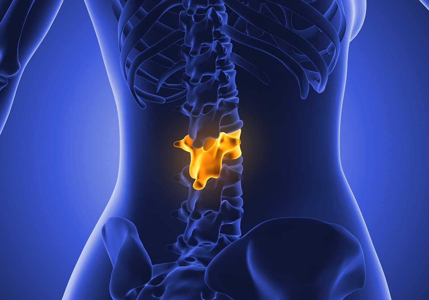
A lumbar disc herniation occurs when the gel-like nucleus pulposus protrudes through a tear or weakness in the outer fibrous ring (annulus fibrosus) of an intervertebral disc. It most commonly affects the L4–L5 and L5–S1 levels of the lumbar spine. The herniated disc material may compress or irritate an adjacent spinal nerve root, causing radiating leg pain, numbness, tingling, or weakness—a syndrome commonly referred to as sciatica when the sciatic nerve distribution is involved. This article follows the patient journey from the onset of symptoms and diagnostic evaluation to treatment options, surgical decision-making, and postoperative recovery.
Quick Answer
- A lumbar disc herniation occurs when the gel-like center of an intervertebral disc (nucleus pulposus) protrudes through a tear or weakness in the outer fibrous ring (annulus fibrosus), most commonly at the L4–L5 or L5–S1 levels.
- The North American Spine Society (NASS) recommends 6–12 weeks of structured nonoperative treatment for most patients, including physical therapy, nonsteroidal anti-inflammatory drugs (NSAIDs) or other appropriate medications, activity modification and/or spinal injections.
- Urgent medical evaluation is required for symptoms suggestive of cauda equina syndrome, including saddle anesthesia, new urinary retention or bowel dysfunction, or for progressive or severe motor weakness in the affected leg.
- Lumbar microdiscectomy removes only the portion of the herniated disc compressing the affected nerve root. Most patients are able to walk on the day of surgery and, depending on recovery and job demands, many return to sedentary work within 1–2 weeks.
- Long-term results from the SPORT (Spine Patient Outcomes Research Trial) demonstrated sustained improvements in leg pain, physical function, and patient satisfaction after surgery, with benefits maintained through 4- and 8-year follow-up. Recurrent disc herniation at the same level occurs in approximately 3%–12% of patients.
What is a lumbar disc herniation, and how does it cause sciatica?
A lumbar disc herniation occurs when the gel-like center of an intervertebral disc (nucleus pulposus) protrudes through a tear or weakened area in the tough outer layer (annulus fibrosus). It most commonly occurs at the L4–L5 and L5–S1 levels of the lower spine. If the herniated disc material compresses or irritates a nearby spinal nerve root, it can cause pain that radiates down the leg—a condition known medically as lumbar radiculopathy and commonly referred to as sciatica.
Each intervertebral disc consists of three main components: the soft, gel-like nucleus pulposus, the strong outer annulus fibrosus, and cartilaginous endplates that anchor the disc to the adjacent vertebrae and help distribute loads. A disc herniation develops when the annulus fibrosus weakens or tears, allowing nucleus pulposus material to bulge outward, extrude through the annulus, or separate completely into the spinal canal (sequestration). Symptoms develop through two primary mechanisms:
- Mechanical compression: Herniated disc material can directly compress a spinal nerve root, producing pain and neurological symptoms.
- Chemical inflammation: The nucleus pulposus releases inflammatory mediators that can irritate the nerve root, causing pain even when mechanical compression is relatively mild.
Sciatica describes pain that radiates along the course of the sciatic nerve, typically from the lower back or buttock down the back or outer side of the leg. Lumbar radiculopathy is the broader medical term for nerve root dysfunction and may cause radiating pain, numbness, tingling, muscle weakness, or diminished reflexes in the characteristic distribution of the affected nerve root. Weakness often follows a predictable myotomal pattern, while sensory changes occur within a corresponding dermatomal distribution.
Importantly, imaging findings must always be interpreted in the clinical context. Disc bulges and even disc herniations are common incidental findings on MRI, particularly with increasing age, and many people with these abnormalities have no back or leg symptoms. For this reason, an MRI finding alone does not establish the cause of pain. A lumbar disc herniation is considered clinically significant only when the imaging findings correspond with the patient’s symptoms and physical examination.
How is a herniated disc diagnosed, and when is an MRI appropriate?
Doctors diagnose a herniated disc by combining three things: a careful history, a focused nerve exam, and an MRI of the lower back when symptoms persist or when warning signs appear. Most early back or leg pain gets better on its own, so routine MRI early on is not recommended.
- Helpful clues in your history: leg pain worse than back pain, pain that follows a specific stripe down the leg, and a clear link to bending, lifting, or twisting.
- Exam clues: a positive straight leg raise test, numbness in a predictable area, weakness in a specific muscle group, and uneven reflexes. The more these signs line up with one spinal level, the more confident the diagnosis.
- An MRI is the best imaging test. It is usually ordered after about six weeks of symptoms that are not getting better, when warning signs are present, or when surgery is being considered.
- A CT myelogram is used for people who cannot have an MRI. Nerve tests (EMG) can help when the diagnosis is unclear.
- An MRI is most useful when it is timed to answer a specific question, not when it is ordered at the very first episode of pain.
Obtaining imaging too early in patients with mild, uncomplicated symptoms may identify incidental abnormalities that are unrelated to the patient’s pain, potentially leading to unnecessary tests or treatments. Conversely, delaying imaging in the presence of red-flag symptoms or progressive neurological deficits can postpone diagnosis and treatment, increasing the risk of permanent nerve injury. For this reason, the clinical history and neurological examination guide the decision to obtain imaging. The exam drives the decision.
What conservative treatments work, and how long should patients try them?
Approximately 60%–90% of patients with a lumbar disc herniation improve without surgery. In the absence of cauda equina syndrome, progressive neurological deficits, or severe motor weakness, the North American Spine Society (NASS) recommends 6–12 weeks of structured nonoperative treatment before considering surgery for most patients. This period reflects the natural history of lumbar disc herniation, as symptoms often improve with time and appropriate conservative care. A comprehensive nonoperative treatment plan typically includes:
- Activity modification: Avoid movements and positions that worsen symptoms while remaining as active as tolerated. Prolonged bed rest is generally discouraged because it can delay recovery.
- Nonsteroidal anti-inflammatory drugs (NSAIDs): NSAIDs and other appropriate analgesics help reduce pain and inflammation, allowing patients to participate more effectively in rehabilitation.
- Physical therapy: A supervised rehabilitation program focusing on core stabilization, flexibility, posture, and gradual return to normal activities is a cornerstone of treatment.
- Oral corticosteroids: A short course of oral corticosteroids may provide modest short-term relief in selected patients with acute lumbar radiculopathy, although benefits are generally limited and should be weighed against potential adverse effects.
- Epidural steroid injections: For patients with persistent or severe radicular leg pain that limits daily activities or participation in physical therapy, epidural steroid injections may provide temporary symptom relief by reducing inflammation around the affected nerve root. These injections relieve symptoms but do not repair or reverse the disc herniation.
Although many patients recover without surgery, conservative treatment should not continue indefinitely when meaningful improvement fails to occur. Therefore, the recommended 6–12-week trial of conservative care represents active treatment with close clinical follow-up, not passive observation. During this period, clinicians monitor symptom progression, neurological function, and response to therapy, while reassessing the need for imaging or surgical referral if recovery plateaus or neurological deficits develop.
When does a herniated disc need surgery?
Most people with a lumbar disc herniation recover without surgery. Surgery is generally recommended in two situations: an emergency, such as cauda equina syndrome or progressive severe leg weakness, or persistent, disabling leg pain that continues after 6–12 weeks of appropriate nonoperative treatment. Before surgery is considered, the patient’s symptoms, neurological examination, and MRI findings should all identify the same affected nerve root.
Urgent surgical evaluation is needed for:
- Cauda equina syndrome: Saddle numbness, new urinary retention or incontinence, bowel dysfunction, or symptoms affecting both legs.
- Progressive or severe motor weakness: Delaying treatment may increase the risk of permanent nerve injury.
Surgery may also be appropriate for persistent sciatica that limits work, daily activities, or quality of life despite conservative treatment, or for recurrent disc herniation causing recurrent nerve compression.
Surgery is not recommended for an incidental MRI finding without matching symptoms, for isolated low back pain without significant nerve root compression, or for symptoms that have been present only a short time in the absence of neurological deficits or other warning signs.
The table below shows how different situations map to surgery urgency.
| Clinical Scenario | Surgical Urgency | Typical Action | Source |
|---|---|---|---|
| Cauda equina syndrome | Emergent | Same-day surgical evaluation | NASS |
| Progressive or severe motor weakness | Urgent | Prompt surgical consultation within days | NASS |
| Persistent radiculopathy after 6–12 weeks of conservative care | Elective | Discuss microdiscectomy with surgeon | NASS, SPORT |
| Imaging finding without concordant symptoms | Not indicated | Continue observation or non-operative care | NASS |
Emergencies and urgent cases are different from elective decisions. Suspected cauda equina syndrome needs immediate emergency care. Fast-progressing weakness needs a surgical consult within days. The elective conversation weighs faster relief against the risks of an operation.
Seek Emergency Care Immediately If You Have:
- Numbness in the groin or inner thighs (saddle anesthesia)
- New trouble urinating or new urinary incontinence
- New bowel problems or loss of bowel control
- Symptoms in both legs at the same time
- Weakness that is getting worse quickly
Do not wait for an office appointment. Go to the nearest emergency room.
What happens during a lumbar microdiscectomy?
A lumbar microdiscectomy is the standard surgical treatment for a symptomatic lumbar disc herniation that does not improve with conservative care or causes significant neurological deficits. It is usually performed as an outpatient or same-day procedure. Through a small incision in the lower back, the surgeon uses an operating microscope or tubular retractor to access the affected nerve root and remove only the portion of the herniated disc compressing the nerve, while preserving the remaining disc, surrounding bone, and spinal stability whenever possible.
The goal of lumbar microdiscectomy is targeted nerve decompression with minimal disruption. The surgeon removes only the fragment of herniated disc that is compressing the nerve root, relieves the pressure causing symptoms, and preserves as much of the normal disc, bone, and spinal structures as possible. Because the procedure is focused and minimally invasive, most patients are able to walk the same day after surgery.
What does recovery look like after microdiscectomy?
Most patients recover quickly after a lumbar microdiscectomy. Many are able to walk within a few hours and go home the same day once they can move safely, control pain with oral medication, and meet standard discharge criteria. During the first 1–2 weeks, recovery focuses on:
- A gradual walking program to restore mobility and endurance
- Incision care and monitoring for signs of infection
- Avoiding heavy lifting, prolonged sitting, and excessive bending or twisting
Many patients return to desk work within 1–2 weeks, depending on symptoms and job demands. More physically demanding work usually requires a longer, gradual return, often around 4–12 weeks, with attention to proper lifting and movement mechanics.
Formal physical therapy typically begins between 2 and 6 weeks after surgery and focuses on rebuilding strength, improving conditioning, and reinforcing safe habits for sitting, lifting, and daily activities. The relatively rapid recovery compared with larger spine operations is one of the major advantages of microdiscectomy.
Call your surgical team if you notice:
- Growing redness or drainage at the incision site
- Fever
- New or worsening leg weakness
- Loss of bowel or bladder control
- Sudden severe leg pain returning
What are the outcomes and long-term results of lumbar microdiscectomy?
Lumbar microdiscectomy provides lasting relief of leg pain and improvement in function for most appropriately selected patients. Key long-term findings include:
- Leg pain improves more predictably than back pain. Microdiscectomy is most effective when symptoms are caused by nerve compression, such as radicular pain that matches the MRI and neurological examination. Patients with primarily low back pain should expect more limited improvement.
- Recurrent disc herniation can occur. Approximately 3%–12% of patients experience another herniation at the same level.
- Risk factors for recurrence may include smoking, obesity, diabetes, older age, and male sex.
- Factors linked to better outcomes include shorter symptom duration before surgery, lower disability levels before surgery, avoiding tobacco use, and addressing significant psychological factors that may affect recovery.
Overall, microdiscectomy has one of the strongest evidence bases among elective spine procedures, with consistently high patient satisfaction in published studies. Long-term results are best when surgery is performed for persistent nerve-related leg pain that matches the clinical findings, rather than for an MRI abnormality alone or isolated back pain. Reducing recurrence risk includes maintaining a healthy weight, avoiding smoking, and using proper body mechanics during lifting and daily activities.
Next Steps
If you think you may have a lumbar disc herniation, the first step is an evaluation by a primary care clinician or spine specialist. This visit typically includes a detailed history, a focused neurological examination, and assessment of whether your symptoms match a possible nerve compression. If there are no warning signs or progressive neurological deficits, most patients begin with 6–12 weeks of structured nonoperative treatment, including activity modification, medications when appropriate, and physical therapy. MRI is generally reserved for patients whose symptoms persist, worsen, or who develop concerning findings that require further evaluation.
Some signs need emergency care right away: numbness in the groin area, new trouble urinating or new incontinence, bowel problems, symptoms in both legs, or weakness that is getting worse quickly. If you have any of these, go to the nearest emergency room immediately. Do not wait for an office appointment.
A spine surgery consultation involves reviewing your symptoms, neurological examination, MRI findings, and treatment goals. A surgical visit does not automatically mean surgery is needed. Depending on your situation, the best plan may be continued nonoperative care, a targeted injection, rehabilitation, or surgery if nerve compression is causing persistent disabling symptoms or neurological problems.
Disclaimer: This article is for educational purposes only and does not constitute medical advice. Every treatment decision is highly individualized. Consult a qualified spine surgeon to determine the appropriate treatment for your condition.

Medically reviewed by
Samia Ghaffar, MD
Board-Certified Orthopaedic Spine Surgeon