

Lumbar spinal stenosis is a condition in which the spaces within the lower spine narrow, reducing the room available for the spinal nerves. This narrowing can compress the nerves, causing leg pain, numbness, tingling, cramping, or a feeling of heaviness, among other symptoms. It is one of the most common degenerative spine conditions in adults over age 60 and a frequent reason for referral to a spine specialist. This article explains how lumbar spinal stenosis is diagnosed, the role of nonoperative treatment, what a lumbar laminectomy involves, and how surgeons determine whether decompression alone is sufficient or whether decompression with spinal fusion is recommended.
Quick Answer
- Lumbar spinal stenosis is a narrowing of the spinal canal or nerve passageways in the lower back that compresses the spinal nerves. It is most commonly caused by age-related degenerative changes, including ligamentum flavum thickening, facet joint hypertrophy, and intervertebral disc bulging.
- The hallmark symptom is neurogenic claudication — leg pain, numbness, cramping, or heaviness that worsens with standing or walking and improves with sitting or leaning forward, often called the "shopping cart sign."
- Initial treatment is usually nonoperative, including activity modification, physical therapy, nonsteroidal anti-inflammatory drugs (NSAIDs) or other appropriate medications, and, for selected patients, spinal injections. Surgery is considered when symptoms remain disabling despite conservative treatment or when neurological function progressively declines.
- Lumbar laminectomy: A lumbar laminectomy relieves pressure on the nerves by removing part of the lamina and thickened ligamentum flavum, enlarging the spinal canal. It only removes a small portion without causing instability.
- Spinal fusion: Spinal fusion is generally reserved for patients with spinal instability, such as symptomatic degenerative spondylolisthesis with instability on flexion–extension X-rays, or for those with significant foraminal stenosis requiring decompression that would otherwise compromise spinal stability.
- Carefully selected older patients, even those in their 70s and 80s, can do well after a laminectomy. A frailty assessment is recommended for patients over 75.
Two surgical options are used to treat these conditions:
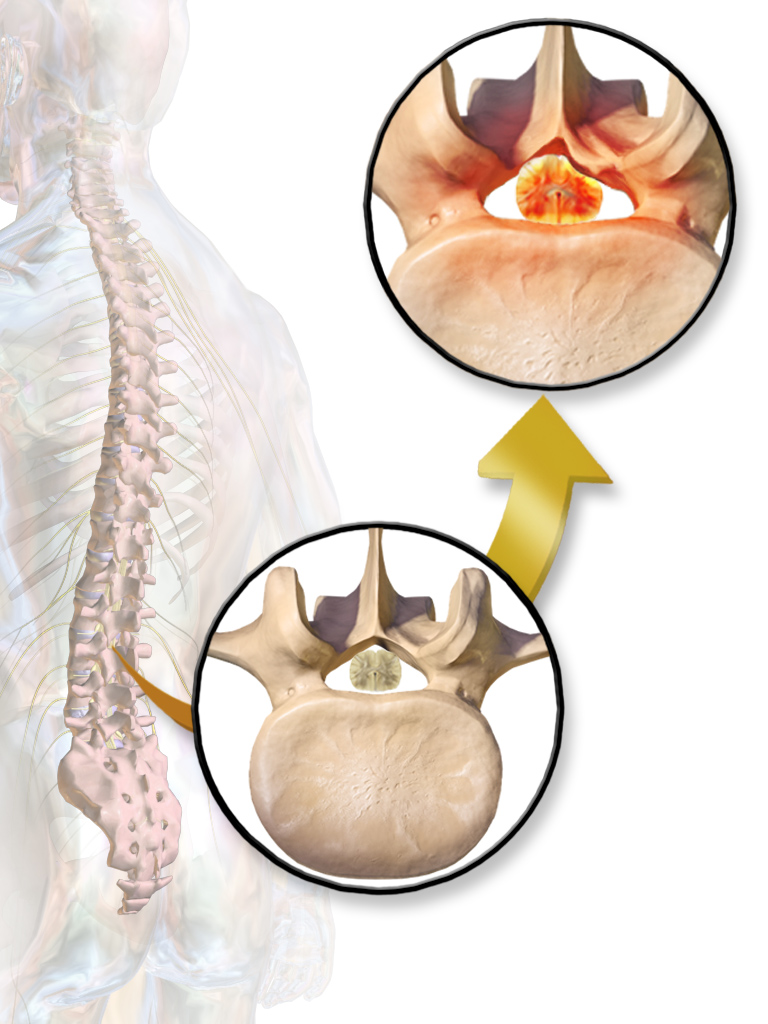
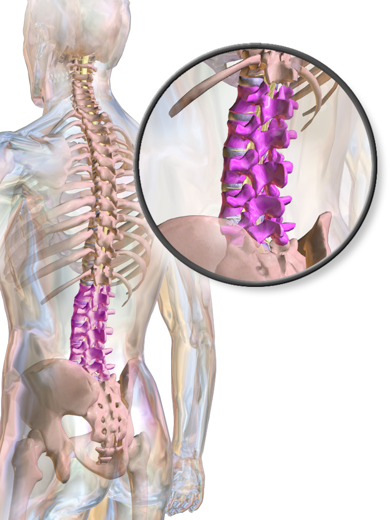
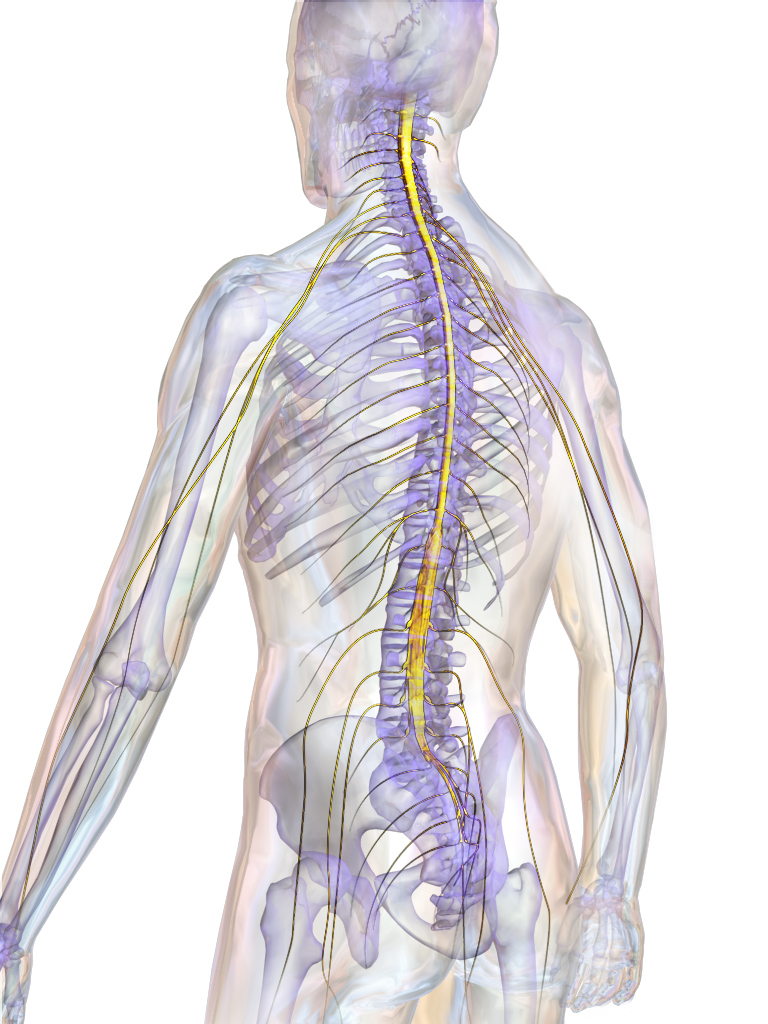
What is lumbar spinal stenosis and why does it make walking harder?
Lumbar spinal stenosis is usually an age-related narrowing of the spaces in the lower spine where the nerves travel. It can be caused by infection, tumor, trauma as well. As these spaces become smaller, the spinal nerves are compressed. Several age-related changes contribute to spinal stenosis:
- Ligamentum flavum thickening: The ligament can thicken and thus reduces the space available.
- Facet joint arthritis: Enlarged facet joints and bone spurs (osteophytes) can narrow the lateral recess and neural foramen, where spinal nerves travel and exit the spine.
- Disc degeneration: Bulging discs and loss of disc height can further narrow the spinal canal and nerve passageways.
The hallmark symptom is neurogenic claudication—pain, heaviness, or fatigue in the buttocks and legs that develops with standing or walking and improves with sitting or bending forward. This relief, often called the "shopping cart sign," occurs because flexing the lumbar spine increases the space available for the nerves. Many patients also find it easier to walk uphill or ride a stationary bicycle than to walk on level ground. MRI findings must be interpreted alongside the patient's symptoms and physical examination to determine whether spinal stenosis is the true cause of the pain.
- The ligamentum flavum can thicken and buckle inward with age, leaving less room for the cauda equina (the bundle of nerves at the bottom of the spinal cord), especially when you stand tall.
- The facet joints can grow bone spurs (osteophytes) and thicken, crowding the lateral recess and the neural foramen where nerves exit.
- Discs can bulge or lose height, pushing into the nerve space.
- Narrowing of the central canal pinches the cauda equina. Narrowing of the lateral recess pinches a passing nerve. Narrowing of the neural foramen pinches the exiting nerve.
- The leg pain or heaviness usually shows up above the knee or in the buttock and thigh. Sitting or leaning forward helps.
- Leg pain from poor blood flow (vascular claudication) usually shows up in the calf, gets better with standing still, and doesn't improve when you bend forward.
- The "shopping cart sign" is the relief people feel leaning forward on a grocery cart, riding a stationary bike, or walking uphill — those positions open the nerve space.
The L4-L5 level is the most common spot. Most people have a mix of narrowing in the central canal, the lateral recess, and the neural foramen. An MRI has to be matched with your symptoms to find the real source of pain — walking trouble has two components: bending forward both opens the space and improves blood flow to nerves that get slightly starved during walking.
How is lumbar spinal stenosis diagnosed?
Lumbar spinal stenosis is diagnosed by combining a careful medical history, a focused physical examination, and MRI findings. The diagnosis depends on matching the patient's symptoms with imaging, as many adults may have spinal stenosis on MRI without experiencing any associated symptoms. The medical history is often the most important clue. Patients typically report leg pain, numbness, cramping, or heaviness that worsens with standing or walking and improves with sitting or leaning forward. Walking tolerance often decreases gradually over months or years. The physical examination may be normal while the patient is at rest, even when symptoms are significant during walking. In more advanced cases, weakness, loss of sensation, or reduced reflexes may be present. MRI is the imaging study of choice because it clearly shows narrowing of the spinal canal, ligamentum flavum thickening, facet joint enlargement, disc degeneration, and compression of the nerve roots. CT myelography is typically reserved for patients who cannot undergo MRI. Because degenerative changes are common with aging, MRI findings alone do not establish the diagnosis. The imaging abnormalities should correspond with the patient's symptoms and physical examination before treatment decisions are made. Electromyography (EMG) and nerve conduction studies are not routinely required but may be helpful when the diagnosis is uncertain or when it is necessary to distinguish lumbar spinal stenosis from peripheral neuropathy or another nerve disorder.
- Your history usually shows position-related leg symptoms, with walking distance that has shrunk over months or years.
- Your exam at rest is often normal — normal strength, reflexes, and feeling — even when you have trouble walking.
- Mild weakness, slower reflexes, or numb spots can show up in more advanced cases. Your doctor will also check walking, balance, hips, and circulation to rule out other causes.
- MRI is the best scan because it shows the ligaments, discs, joints, and fluid around the nerves.
- CT myelogram is saved for people who can't have an MRI.
- Many older adults have stenosis on imaging without symptoms, so the scan has to match the story.
- EMG and nerve conduction studies are not routine — they can help when it's hard to tell stenosis from a neuropathy or a single pinched nerve.
What are conservative care modalities, and when is it enough?
For most patients, the first step is nonoperative treatment. A typical trial lasts 6–12 weeks and aims to reduce symptoms, improve walking tolerance, and maintain function. A comprehensive treatment plan typically includes:
- Activity modification: Stay as active as possible while avoiding prolonged activity that can trigger symptoms.
- Physical therapy: A supervised program focuses on core and hip strengthening, stretching, balance training, and gait retraining. Therapists also teach posture and body mechanics that help reduce pressure on the spinal nerves.
- Medications: Nonsteroidal anti-inflammatory drugs (NSAIDs) or other pain relievers may help control symptoms and improve participation in physical therapy. In selected patients, medications for nerve-related pain may also be considered.
- Epidural steroid injections: For patients with persistent leg pain that limits walking or participation in rehabilitation, an epidural steroid injection may provide temporary relief by reducing inflammation around the compressed nerves. These injections can improve function but do not reverse the underlying spinal narrowing.
Conservative treatment is considered successful if leg symptoms improve, walking distance becomes acceptable, daily activities can be performed with manageable discomfort, and there is no progressive neurological deficit.
Seek Emergency Care Immediately If You Have:
- New leg weakness
- New bladder or bowel problems, including urinary retention or incontinence
- Numbness in the groin or inner thighs (saddle anesthesia)
These can signal cauda equina syndrome — a rare but serious medical emergency. Do not wait for an office appointment. Go to the nearest emergency room.
What is a lumbar laminectomy?
A lumbar laminectomy is one of the standard surgical treatments for symptomatic lumbar spinal stenosis. During the procedure, the surgeon removes part of the lamina (the back portion of the vertebra), along with the thickened ligamentum flavum and any bone spurs or other tissue compressing the nerves. The goal is to decompress the spinal canal, giving the nerves more room and relieving leg pain and neurogenic claudication while preserving as much of the normal spinal anatomy as possible.
- A hemilaminectomy is a laminectomy on only one side, used when pinching is mostly on one side.
- A laminotomy is a smaller version, where less bone is removed.
- A minimally invasive approach works through a small tube (a tubular retractor) instead of a long cut. Minimally invasive and traditional open approaches give similar symptom results; minimally invasive has less blood loss and shorter stays.
- Risks include bleeding, infection, a small tear in the nerve lining with fluid leak, blood clots, anesthesia problems, and a small chance of a second operation.
- Risk rises with more levels, with fusion, and with frailty — so early walking and close follow-up matter.
When is fusion necessary?
A spinal fusion is not needed for some patients undergoing surgery for lumbar spinal stenosis. It is generally reserved for situations in which the spine is unstable or is expected to become unstable after decompression. Fusion may be recommended for patients with:
- Degenerative spondylolisthesis (one vertebra slipping over another) with evidence of instability on flexion–extension X-rays.
- Significant spinal deformity, such as scoliosis.
- Severe foraminal stenosis requiring extensive bone removal that would compromise spinal stability.
- Recurrent stenosis or prior surgery in selected cases.
The decision to add a fusion is based on each patient's anatomy, imaging findings, and symptoms. The goal is to relieve nerve compression while preserving spinal stability, using the least invasive procedure that is likely to provide a durable result.
- The SLIP trial (NEJM 2016, 66 patients) — at 2 years, the fusion group had a small but real edge on quality-of-life score (SF-36 PCS) and fewer needed a second operation.
- The Försth trial (Swedish NEJM 2016, 247 patients) — at 2 and 5 years, the fusion group didn't do better on patient-reported outcomes. Operations were longer, with more bleeding and longer stays.
- NORDSTEN-DS (Norwegian NEJM 2021, 267 patients) — decompression alone was just as good as decompression plus fusion at 2 years on the Oswestry Disability Index.
- NORDSTEN-DS 5-year follow-up (Lancet 2024) — confirmed decompression alone held up just as well long-term.
- Newer reviews lean toward decompression alone for most mildly slipped bones. Patients with clear instability or a curve still benefit from fusion.
What does recovery from a lumbar laminectomy look like?
Recovery after a lumbar laminectomy without fusion is generally faster than recovery after fusion surgery. Most patients walk on the day of surgery, go home the same day, and gradually return to normal activities over the following weeks. Recovery after laminectomy with fusion is longer because the bones must heal together, with a hospital stay of 2-3 days and full recovery often taking 3–6 months. During the first few weeks after surgery:
- Walking is encouraged immediately and is the best way to rebuild strength and endurance. Increasing walking distance is one of the most useful measures of recovery.
- Pain is typically managed with a short course of prescription pain medication, followed by acetaminophen and, when appropriate, anti-inflammatory medications.
- Heavy lifting, repetitive bending, and twisting should be avoided until cleared by your surgeon. These restrictions generally last longer after fusion surgery.
- Most patients can resume driving once they are no longer taking opioid pain medication and can safely perform an emergency stop, often 2–4 weeks after decompression alone.
- Return to desk work is common within 2–6 weeks, while physically demanding jobs may require several months and a gradual return-to-work plan.
- A frailty assessment is recommended for patients over 75. Carefully chosen older patients can have a real boost in quality of life.
- The SPORT trial showed real surgical benefit at 2 and 4 years. At 8 years, the observational cohort continued to show a surgical advantage. Leg symptoms usually improve more reliably than back pain.
How do patients and surgeons decide?
Choosing between continued nonoperative care, laminectomy alone, or laminectomy with fusion is a shared decision based on the patient's symptoms, imaging findings, overall health, and treatment goals. Key factors include:
- Symptom severity: Are leg pain and walking limitations significantly affecting daily life?
- Response to conservative treatment: Has an adequate 6–12-week trial of nonoperative care failed to provide meaningful relief?
- Spinal stability: Do flexion–extension X-rays show instability, or would decompression alone leave the spine unstable?
- Overall health: Age, medical conditions, and frailty influence surgical risk and recovery.
- Symptom pattern: Surgery is generally more effective for leg pain and neurogenic claudication than for isolated low back pain.
For many patients with a stable spine, a laminectomy alone provides excellent symptom relief while avoiding the added recovery and risks of fusion. Fusion is usually reserved for patients with documented instability or when the decompression required to relieve nerve compression would compromise spinal stability.
Next Steps
A few simple steps can make your spine visit more productive. Track your activity level and daily limitations over time, including how far you can walk, what activities trigger symptoms (such as standing, flat ground, hills, or grocery aisles), and what provides relief. Bring prior imaging and reports, a current medication list, and a summary of previous treatments, including physical therapy, injections, medications, and any prior spine surgeries with dates and outcomes.
Seek urgent care for new leg weakness, new bladder or bowel problems, or numbness in the groin or inner thighs — those can signal cauda equina syndrome, which is a surgical emergency.
Disclaimer: This article is for educational purposes only and does not constitute medical advice. Consult a qualified spine specialist for evaluation and diagnosis of your specific symptoms.
Medically reviewed by
Samia Ghaffar, MD
Board-Certified Orthopaedic Spine Surgeon